An open, well-shaped eye.
"Epicanthoplasty is as much about the shape and proportions of the eye as it is about the size. Done well, it produces a clearer eye line and a softer overall expression."
— Director Choi Dong-Il
Both the shape and the size of the eye, addressed in a single procedure.
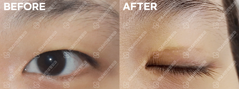
When the eye still looks narrow despite a natural or surgically created fold, extending its horizontal length opens up the appearance and produces a clearer, more defined eye line.
The same procedure refines both the size and the shape of the eye, so the result can be tailored to what each patient is looking for.
※ Surgical and recovery details may vary depending on the patient's individual condition.
From surgery time to return to daily activity, all in one view.
Five clinical situations in which epicanthoplasty is the appropriate choice.
A defined fold,
but the eye still looks narrow
Short horizontal length
making the eye look small
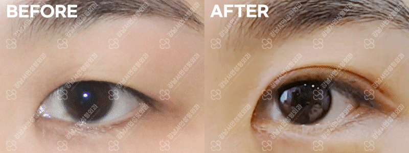
Outer corners that
droop or tilt upward
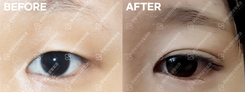
Prominent
epicanthal folds
Patients seeking a
larger, more open eye
Epicanthoplasty has become a popular procedure, but performed without regard to the patient's actual anatomy, it can worsen scleral show or leave the expression looking sharper than intended. A thorough consultation with an experienced plastic surgeon is essential before deciding whether to proceed.
Director Choi Dong-Il, a board-certified plastic surgeon, personally evaluates indication, performs the surgery, and oversees follow-up.
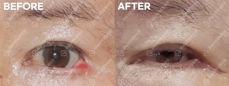
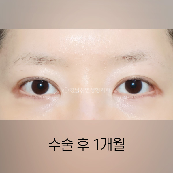
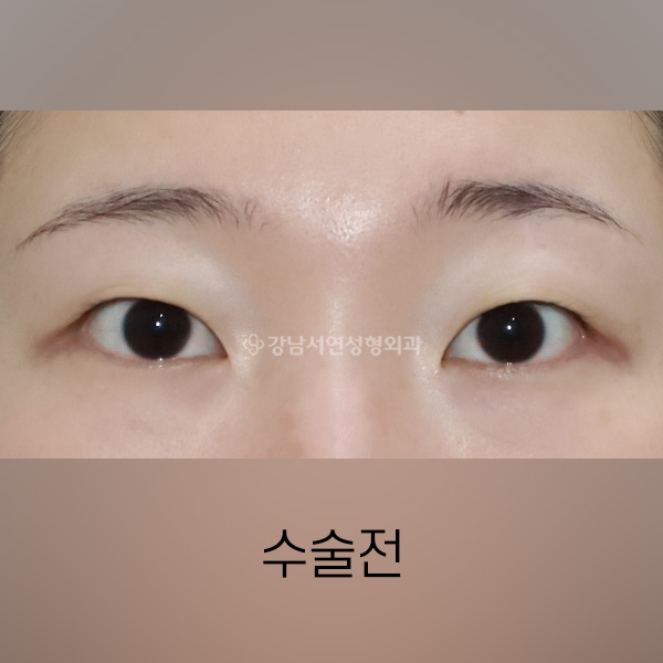
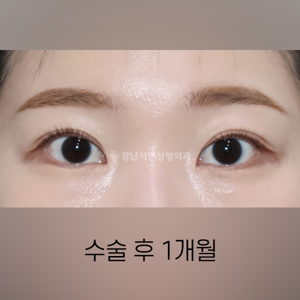
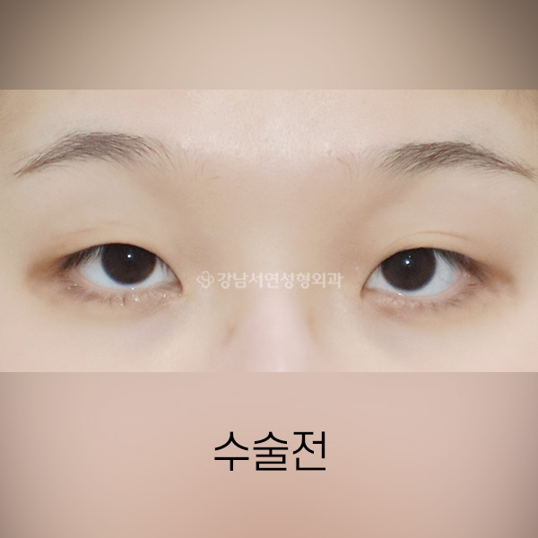
We assess the strength of the epicanthal fold, the proportions of the eye, and how much of the caruncle (the pink tissue at the inner corner) is currently exposed.
Each technique is individualized in design and execution to produce a natural-looking result.
Skin redraping technique — a scar that becomes nearly invisible over time.
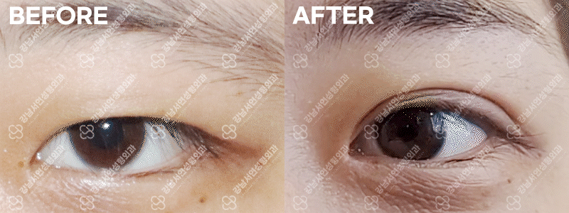
We assess the shape of the fold and the proportions of the eye, along with how much of the caruncle (the pink tissue at the inner corner) is currently visible. Based on that assessment, the design is individualized and the procedure is carried out using the skin redraping technique. We generally aim for 70–80% caruncle exposure as the most balanced result (presented at academic conferences in 2016 and 2020). The scar tends to fade until it is nearly invisible over time.
Caruncle hidden behind the epicanthal fold.
Skin redraping just completed.
Stitches removed, in early recovery.
Performed in roughly 30% of cases — only after the eye is confirmed suitable.
We first confirm whether the eye is genuinely suited to lateral or lower canthoplasty before working out the individualized design. In our practice, only about 30% of patients are appropriate candidates — both for a meaningful effect and for a natural-looking result. The eye is opened up gently, with care taken to avoid excessive scleral show. In some cases the lid does not sit perfectly against the eyeball immediately after surgery; this typically resolves within a few days.
Performed alone, lateral or lower canthoplasty often re-adheres or falls short of the expected result; combining the two is generally recommended.
Lower canthoplasty is not recommended for patients with lash-line irritation, fullness under the eye, or scleral show; in those cases, skin excision or another approach is more appropriate.
Published in Plastic and Reconstructive Surgery (2019) — a Gangnam Seoyon technique.
When too much caruncle has been exposed after a previous medial epicanthoplasty, medial epicanthoplasty reconstruction can correct the appearance. We restore the inner corner while keeping the original scar as inconspicuous as possible.
Our medial epicanthoplasty reconstruction technique was published in the American journal Plastic and Reconstructive Surgery.
Reserved for case GIFs of medial epicanthoplasty reconstruction.
A patient who had been left with excessive caruncle exposure after a previous epicanthoplasty. This is the unedited one-year follow-up.
The lateral canthal tendon refixed to bone or periosteum.
When a previous lateral canthoplasty has left too much conjunctiva exposed — leaving the outer corner looking downturned, or causing functional issues — reconstruction is needed. The end of the lateral canthal tendon is fixed back to bone or periosteum to rebuild the corner.
Excessive conjunctival exposure at the outer corner.
The lateral canthal tendon refixed for a natural finish.
The lateral canthal tendon refixed to bone and periosteum. Pre-op, immediate post-op, and the one-week recovery — all in a single video.
Techniques validated through academic presentations and live surgical demonstrations.
Presentation on medial epicanthoplasty reconstruction and epicanthal fold restoration.
Live surgery lecture on incisional ptosis correction and medial epicanthoplasty.
Four outcomes patients can expect from epicanthoplasty.
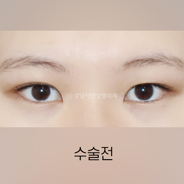
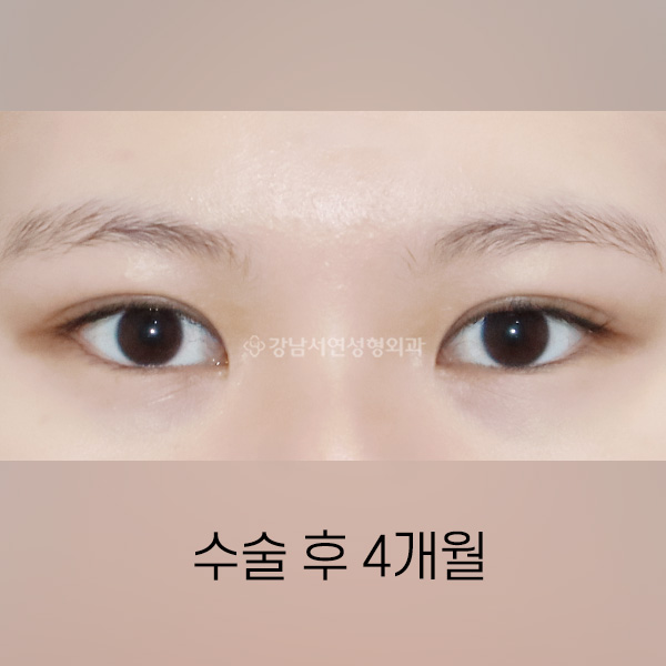
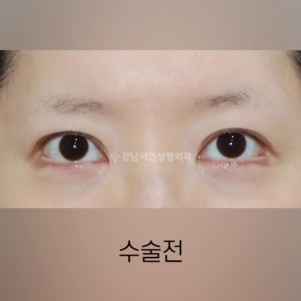
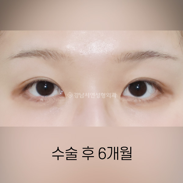
All photographs shown are taken six months after surgery. Additional cases are available in the Before & After section of the menu.
Long-form videos walking through epicanthoplasty, presented by Dr. Choi.
Every inquiry is reviewed personally by Director Choi Dong-Il.