Quick ConsultHoursDirectionsAll Menus
EYELID SURGERY

Incisional Double Eyelid

One precise design. A clean fold that lasts.

"

One precise design,
built to hold its shape over time.

"Incisional surgery does not have to leave a visible scar. With careful design, the orbicularis oculi preserved, and a clean closure, the scar typically becomes very inconspicuous, and the fold tends to outlast every other technique."

— Director Choi Dong-Il

ABOUT

What Is the Incisional Method?

Reducing thick skin and excess fat to create a defined fold that holds its shape over time.

The Most Durable Approach to Double Eyelid Surgery

The incisional method opens the eyelid along the planned design line and shapes the fold while reducing thick skin, excess fat, and connective tissue. The line is set precisely in a single procedure and tends to remain stable longer than any other double-eyelid technique.

It is the appropriate choice for thicker eyelids, sagging skin, and folds that have come undone after a non-incisional procedure. Two factors are decisive in the final result: a clean surgical closure and preservation of the orbicularis oculi, the muscle responsible for closing the eye.

※ Surgical and recovery details may vary depending on the patient's individual condition.

VISUAL · GIF
Incisional method — animated walkthrough
PROCEDURE INFO

Procedure at a Glance

Candidates · method · result — all in one view.

Surgery Time
40–80 min
Anesthesia
Sedation / Local
Visits
1–2
Stitch Removal
After 5–7 days
Hospitalization
Same-day discharge
Daily Activity
After 5–7 days
Who
Thicker Skin or Sagging Eyelids

Patients with excess fat, or with folds that have come undone after a non-incisional procedure.

How
Precise Incision with Muscle Preserved

Skin, fat, and connective tissue are reduced while the fold takes shape.

Result
A Defined, Long-Lasting Fold

A natural line with the scar tucked into the crease, producing the most durable double-eyelid result.

Surgeon
Director-Led, Start to Finish

Director Choi Dong-Il personally handles consultation, surgery, and follow-up, drawing on twenty-two years of eyelid surgery practice.

CANDIDATES

Suitable Candidates

Five clinical situations in which the incisional method is the appropriate choice.

01

Thicker eyelid skin
or excess fat

02

Folds that have repeatedly
come undone after non-incisional surgery

03

Sagging or aging eyelids
requiring a clearly defined fold

04

Patients seeking
a long-lasting result

05

Patients who want
a precisely designed fold

SAFETY · MISCONCEPTION

Does the incisional method always leave a visible scar?
The answer is NO.

An incision does not necessarily mean a visible scar. With careful design, preservation of the orbicularis oculi (the muscle responsible for closing the eye), and a clean surgical closure, the scar typically becomes very inconspicuous. We evaluate each patient's individual eyelid anatomy and recommend the method that genuinely fits.

Always leaves a scar? NO.
Swelling lasts indefinitely? NO.
Always produces a heavy fold? NO.
Orbicularis Oculi Preserved — Our Signature

Damage to the eye-closing muscle can interfere with tear drainage and leave the eyes unable to close fully. Preserving it during surgery is essential to a healthy long-term result.

Closure Tucked Inside the Fold

Two decades of director-led closures, with the suture line set just inside the fold so that it settles naturally and remains very inconspicuous.

METHOD

Surgical Steps

Skin, fat, and connective tissue are reduced while the orbicularis oculi is preserved.

PROCEDURE STEP

The Incisional Method in Five Steps

Design, incision, tissue reduction, and closure.

A precise incision is made along the design line. Thick skin, excess fat, and connective tissue are then reduced while the fold takes shape. Preservation of the orbicularis oculi — the muscle responsible for closing the eye — is decisive in distinguishing a healthy result from a compromised one, and is among our signature techniques. The fold is set with dynamic fixation so that it does not loosen over time.

STEP 01
📐
DIAGNOSIS
Assessment of the eyelid
and selection of method & design
STEP 02
INCISION
Precise incision along
the individualized design
STEP 03
REDUCE
Reduction of thick skin,
fat, and connective tissue
STEP 04
🧵
SUTURE
Muscle preserved,
secured with dynamic fixation
STEP 05
RESULT
Postoperative result
🎬
CORE · GIF
Orbicularis preservation — our signature
SIGNATURE SKILL

Orbicularis Oculi (Eye-Closing Muscle), Preserved

Damage to this muscle can leave the eye unable to close fully and can compromise tear drainage. Working around the muscle, rather than through it, is essential to a stable, healthy result.

🎬
DETAIL · GIF
Closure tucked inside the fold
CORE TECHNIQUE

Closure Tucked Inside the Fold

The closure is placed just inside the fold so that it settles naturally into the crease. Over the months that follow, it generally becomes very difficult to detect.

CASE GIF

Surgical Cases

Actual surgical footage. Only watermarked clips that protect patient identity are posted.

Semi-outline incisional fold

Semi-Outline Incisional Fold

Incisional fold + medial epicanthoplasty

Incisional Fold + Medial Epicanthoplasty

Incisional fold + medial epicanthoplasty

Incisional Fold + Medial Epicanthoplasty

Incisional fold

Incisional Fold

Incisional fold + medial epicanthoplasty

Incisional Fold + Medial Epicanthoplasty

Incisional fold + medial epicanthoplasty

Incisional Fold + Medial Epicanthoplasty

Upper eyelid drooping correction

Upper Eyelid Drooping Correction

Loose fold revision

Loose Fold Correction (Revision)

Upper-eye fat grafting + incisional revision

Upper-Eye Fat Grafting + Incisional Revision

RESULT

Expected Results

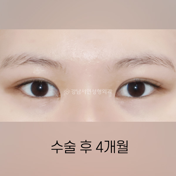
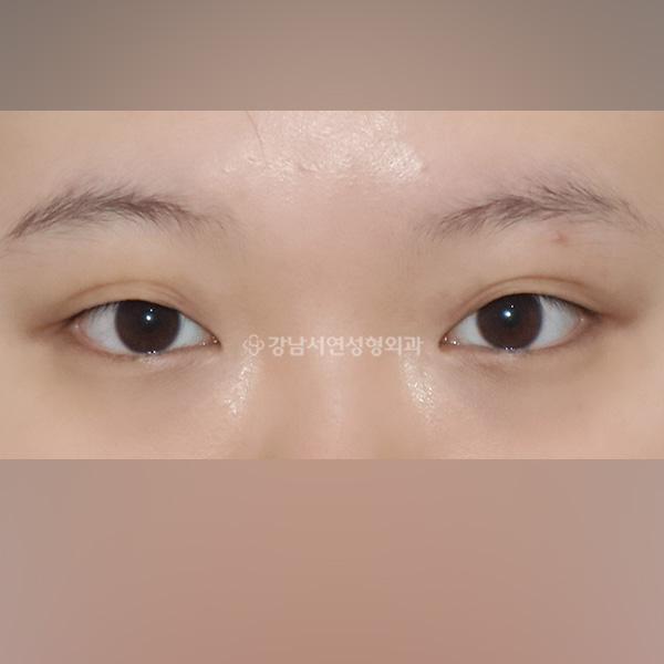
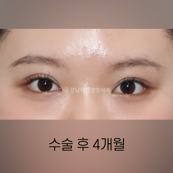
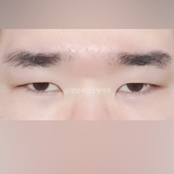
Four outcomes patients can expect from the incisional method.

BEFORE / AFTER · GIF
Animated before and after
Important Information As with any surgery, possible complications include bleeding, infection, and inflammation; severity may vary. Please follow your surgeon's instructions carefully.
BEFORE & AFTER

Patient Results

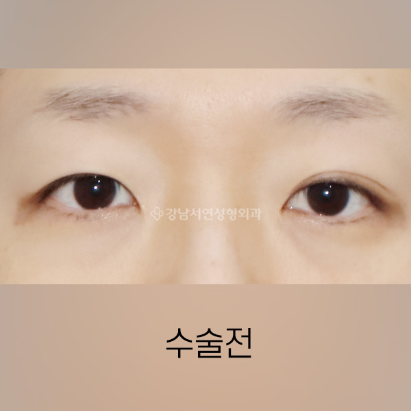
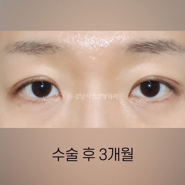
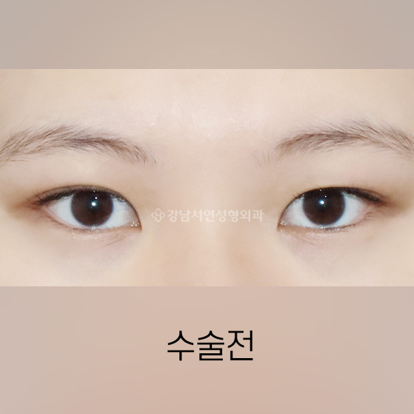
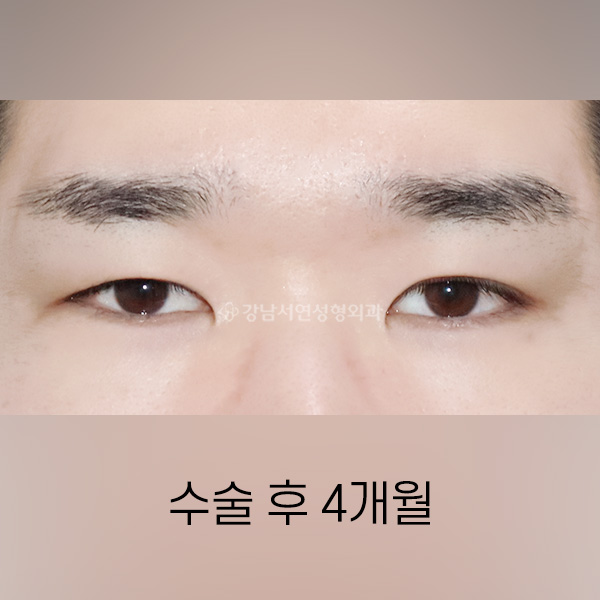
All photographs shown are taken six months after surgery. Additional cases are available in the Before & After section of the menu.

Incisional double eyelid BEFORE Incisional double eyelid AFTER
BEFORE
AFTER
#IncisionalDoubleEyelid

Incisional Double Eyelid

knsy case #13
Incisional + medial epicanthoplasty BEFORE Incisional + medial epicanthoplasty AFTER
BEFORE
AFTER
#IncisionalDoubleEyelid#MedialEpicanthoplasty

Incisional Double Eyelid + Medial Epicanthoplasty

knsy case #8
Semi-outline incisional double eyelid BEFORE Semi-outline incisional double eyelid AFTER
BEFORE
AFTER
#IncisionalDoubleEyelid

Semi-Outline Incisional Double Eyelid

knsy case #71
Incisional + lateral + lower epicanthoplasty BEFORE Incisional + lateral + lower epicanthoplasty AFTER
BEFORE
AFTER
#IncisionalDoubleEyelid#LateralEpicanthoplasty#LowerEpicanthoplasty

Incisional Double Eyelid + Lateral + Lower Epicanthoplasty

knsy case #69
More Before & After
EXPERT GUIDE

Director's Surgical Notes

Long-form videos walking through the incisional method, presented by Dr. Choi.

FAQ

Frequently Asked Questions

Does incisional surgery scar more than the suture method?
When performed correctly, no. The closure is placed just inside the fold and fades into the crease over the months that follow. With the orbicularis oculi preserved and the suture line tucked away, scarring is generally kept to a minimum.
Who is a good candidate for the incisional method?
Patients with thicker eyelid skin, excess fat or connective tissue, sagging eyelids, or folds that have repeatedly come undone after non-incisional surgery. It is also the appropriate choice when a patient wants a precisely designed fold that is built to last.
How long does recovery take?
Stitches are removed at five to seven days, with most of the visible swelling settling within one to two weeks. Most patients return to daily activities within about a week, and the fold typically finishes settling over three to six months.
Can the fold come undone?
Very rarely. Unlike the non-incisional method, the incisional method reshapes the underlying tissue itself, so there is far less to give way. We additionally apply dynamic-fixation suturing for long-term stability.
Can the procedure be combined with epicanthoplasty?
Yes. Adding a medial or lateral epicanthoplasty alongside the incisional method can open the gaze and produce a cleaner, more defined eye shape.
Can ptosis be corrected at the same time?
Yes. During the incisional procedure, the levator aponeurosis can be adjusted to correct ptosis as well, helping to replace a heavy, half-shut appearance with a more open, awake look.
Important Information As with any surgery, possible complications include bleeding, infection, and inflammation. Individual results vary; please consult with your physician for the appropriate diagnosis and treatment plan.
CONSULT

Schedule a Consultation

Every inquiry is reviewed personally by Director Choi Dong-Il.

Clinic
Blog
Doctor's
Blog
Cafe KNSY
TV
Insta-
gram
Kakao
Channel
Doctors Eyes Events Before
& After
Patient
Reviews
B&A
Videos
Kakao
Talk
+82-2
535-8889
Find Us TOP ↑