A case from a few days ago, where we performed both procedures together.
Her crease sits high and reads as a classic sausage fold.
The crease looks high, the fixation looks tight, and the lashes appear lifted.
But measure the line with the eye closed and it actually is not particularly high. So why does it read high?
Because skin is in short supply. And because the scarring is significant.
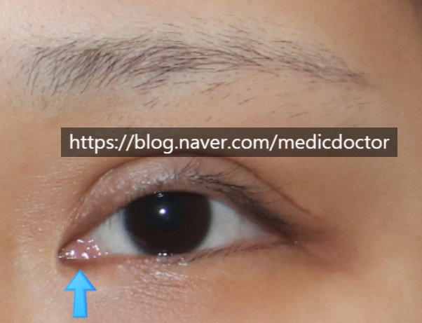
The caruncle is also very visible in front — fully exposed at 100 percent.
The caruncle is the small reddish, clam-shaped tissue at the medial corner.
Excessive medial epicanthoplasty leaves the caruncle 100 percent exposed.
Western patients tend to have no Mongolian fold to begin with.
Quick illustration.

A Western eye normally has the caruncle fully exposed in front. The same look on an Asian face reads as unnatural.
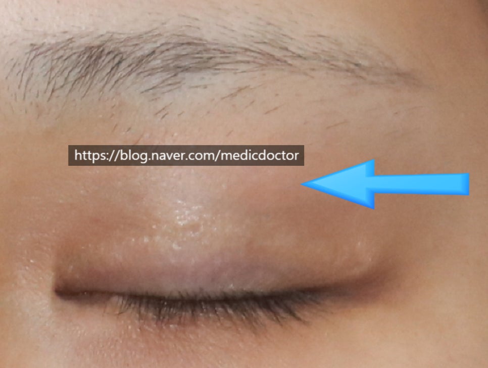
Closed-eye view.
Closed, the crease is set lower than usual. But when I gently pinched the upper-lid skin, there was almost no slack to work with — particularly in the area marked by the arrow.
The plan: set a new crease below the existing one, fully release the adhesion, and prevent re-adhesion. Those are the keys.
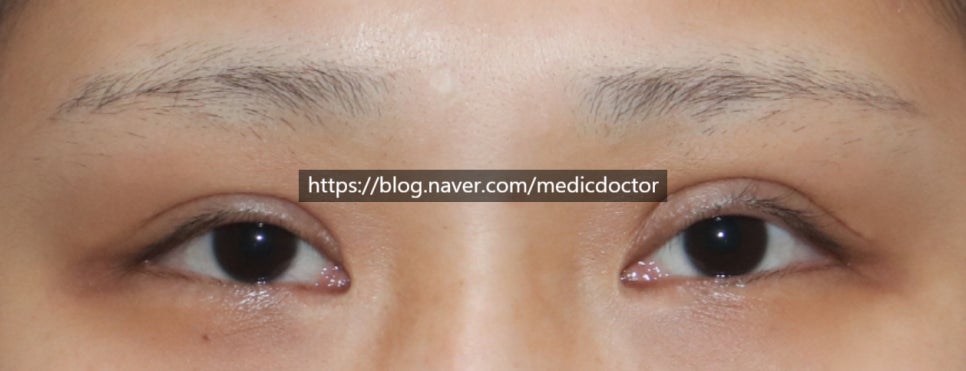
Pre-op view.
The crease is high and asymmetric. One side (her left, the right side of the photo) had a particularly tight, high fixation with more pronounced scarring.
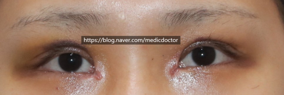
One week post-op, at suture removal. Still swollen, but the line has come down nicely.
The caruncle is also significantly less exposed. Plenty of swelling still to resolve.
For the medial epicanthoplasty reversal, see my 2019 paper.
Her right eye (the left side of the photo) also has some bruising.
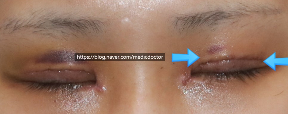
The upper arrow points to the original crease, faintly visible in white.
The lower arrow points to the new crease.
For a medial canthus reversal, the closure should sit slightly raised — it flattens out over time.
More on the two-line revision in other posts.
I tried my hand at video editing here. Still rough.
I hope the swelling resolves cleanly and everything settles in well.