Between conference prep, textbook chapters, and writing papers, my blog has admittedly been quiet lately.
Still, I want to write up one of our recent lateral epicanthoplasty reconstructions.
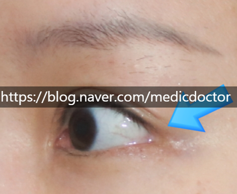
When a lateral canthal release is overdone, the corner of the eye opens into an inverted-U shape and the conjunctival mucosa becomes exposed.
In that situation we perform a revision: the scarred mucosal tissue is removed and the lateral canthal tendon is re-anchored firmly to the lateral orbital periosteum so the deformity does not recur.
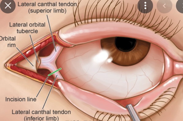
The lateral canthal tendon attaches to the lower eyelid and supports the lid from both sides; it also helps the eye close. When the lateral canthus is over-dissected during the original surgery, the tendon detaches from the periosteum and the corner takes on that inverted-U shape.
So the keys to this surgery are: cleanly excising the scarred mucosa, repositioning the released lateral canthal tendon, preventing recurrence, and hiding the incision well.
The most common patient question is whether the eye will look smaller after surgery.
If the canthus had been excessively opened and we close it considerably, the eye can look slightly smaller. When the issue is mainly the exposed scar, however, the eye does not actually shrink.
The suture-removal marks still look red right now, but they fade to nearly invisible with time.
An older post on this topic is worth reviewing as well.