I want to walk through a case of lateral canthoplasty reconstruction — pre-op, immediately post-op, and one week out.
When a lateral canthoplasty is pushed too aggressively, the lateral canthal tendon can be damaged. The corner then loses its normal contour and takes on a squared, U-shaped appearance.
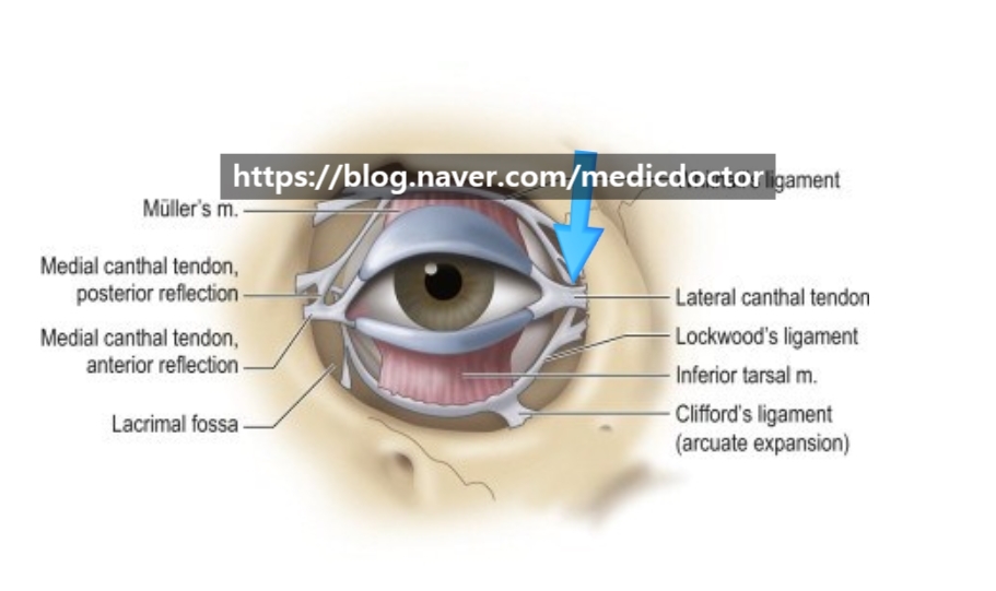
The lateral canthal tendon sits at the position marked by the arrow above.
When this structure is injured, the tendon detaches from its anchor and the conjunctival mucosa becomes exposed.
The eye may also fail to close fully, and the corner can take on a downturned, sad-looking shape.
Here is the comparison: pre-op, and one week post-op at suture removal.
And here is the same case on video.
The reconstruction follows three principles.
1. Cleanly excise the existing scar tissue.
2. Identify the displaced lateral canthal tendon and re-anchor it to its anatomic position.
3. Re-approximate the skin so the scar is as inconspicuous as possible.
Interestingly, the eye does not look smaller afterward — if anything, it looks larger. Maybe that is just my eye for it.
The video tells the story even more clearly than the photos.
Rather than chasing an aggressive lateral opening, a measured release with minimal scarring is the better strategy.
I would much rather a patient have a modest lateral canthoplasty than push too far and end up needing a reconstruction.