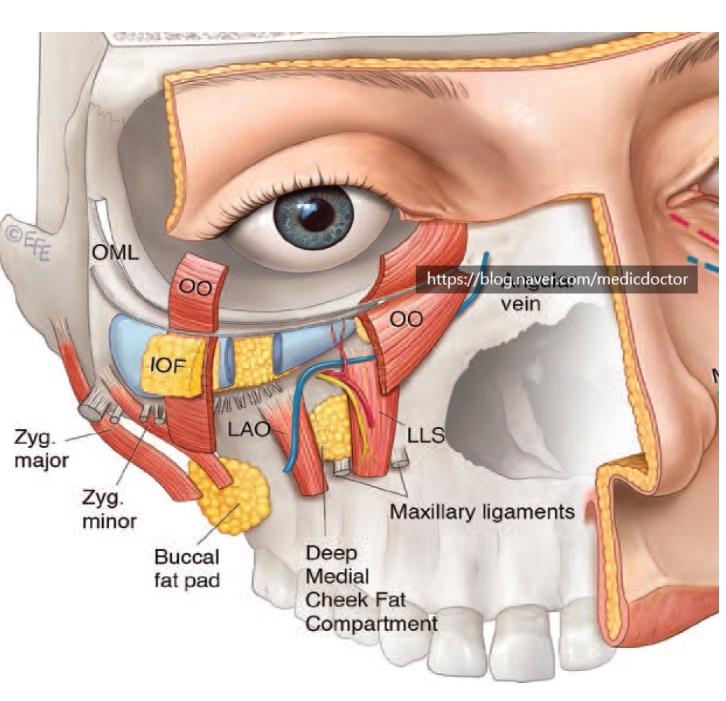
When you perform lower eyelid fat repositioning, a solid grasp of the regional anatomy is what keeps the operation safe
and prevents complications.
I am sharing this for anyone who would find it useful.
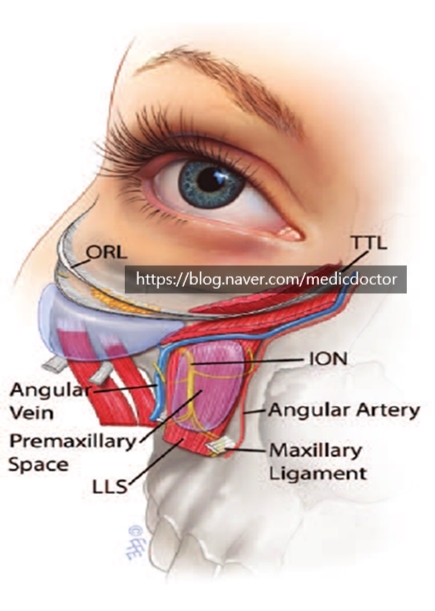
Upper-half roof: orbital part of the orbicularis oculi muscle.
Lower-half roof: mid-cheek SMAS.
Floor: levator labii superioris (LLS).
Superior border: tear trough ligament (TTL).
Inferior border: maxillary ligament (ML).
Medial border: nasal sidewall, levator labii superioris alaeque nasi (LLSA), nasalis.
Lateral border: medial pupil line.
This diagram is well known in the field.
During dissection, you have to watch the angular vein, and the premaxillary space must be released cleanly so the SOOF can be elevated.