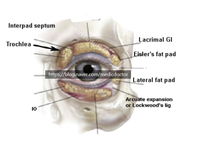
Anatomy of the periorbital fat: orbital fat compartments and the preaponeurotic fat.
Lower eyelid.
Orbital septum.
1. Origin at the arcus marginalis, where the periosteum and periorbita fuse with the orbital rim across a 1-3 mm thickening. Not a single sheet but several thin membranous layers.
2. About 5-6 mm below the inferior tarsal border, the septum joins the capsulopalpebral fascia (CPF).
3. The upper portion is reinforced by the CPF; the lower portion is unreinforced.
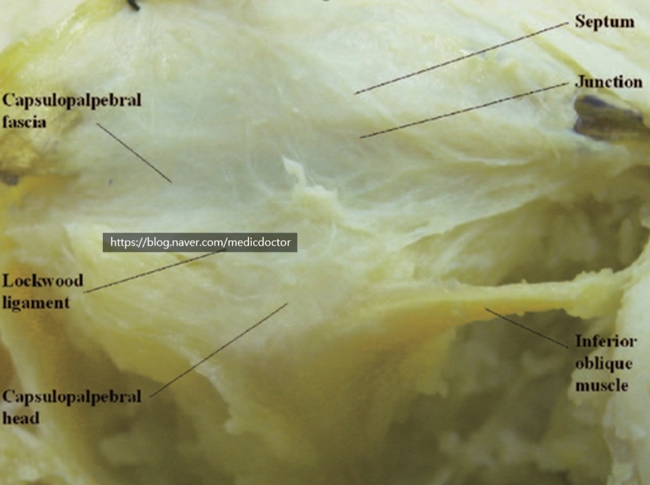
Capsulopalpebral fascia.
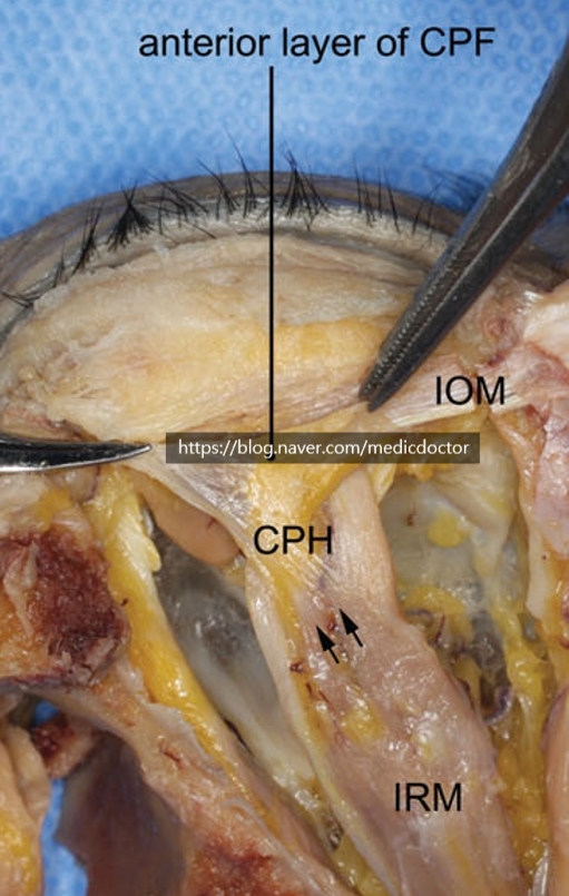
A well-defined connective tissue layer that mechanically links the lower eyelid to the downward retractor apparatus of the globe.
It consists of two distinct layers extending forward and upward from Lockwood's ligament as dense fibrous sheets. The anterior (superficial) layer is coarser and inserts onto the orbital septum and the deep fascia of the orbicularis oculi. The denser posterior layer inserts onto the inferior border of the tarsal plate.
1) Capsulopalpebral head (CPH): originates from the inferior rectus muscle fascia, wraps around the inferior oblique, and reaches Lockwood's ligament. About 7 mm thick.
2) CPF: courses from Lockwood's ligament to the lower margin of the tarsus and the subcutaneous tissue.
3) White, glistening, two-layered structure: a thin anterior layer and a thicker posterior layer.
Arcuate expansion.
1) A fibrous band extending from the infraorbital rim to the medial canthal tendon.
Fan-shaped, tapering to about 2-3 mm in width before its insertion at the medial canthal tendon.
2) Lies deep to the orbital septum and superficial to the inferior oblique muscle.
3) Crosses the middle portion of the inferior oblique and connects with the CPF.
4) Lockwood's ligament and the arcuate expansion originate medially at the posterior lacrimal crest and partition the central from the lateral fat pad.